
Moms-to-be could soon watch babies grow in the womb on a smartphone.
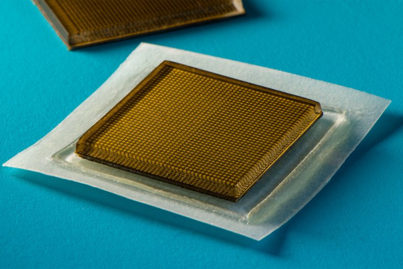
Postage stamp-sized adhesive patches have been developed to provide imaging of the heart, lungs, and other organs.
They will be connected to your smartphone – producing clear, continuous images for 48 hours.
While women could see their own fetuses, it would also improve tumor tracking for cancer patients.
They have a host of potential applications for accelerating disease diagnosis and treatment.
Jon Mills, SWNS/Zenger
“We envision a few patches stuck to different places on the body, and the patches would communicate with your mobile phone, where AI algorithms would analyze the images on demand,” said study lead author Professor Xuanhe Zhao. , a mechanical engineer at the Massachusetts Institute. of Technology.
“We think we’ve ushered in a new era of wearable imaging. With a few patches on your body, you could see your internal organs.”
The Boston-based institution’s team carried out a battery of tests with healthy volunteers, who wore the stickers on various parts of their bodies, including the neck, chest, abdomen and arms.
They remained attached to their skin and took detailed pictures of the underlying structures for up to two days.
During this time, participants performed a variety of activities in the lab ranging from sitting and standing to jogging, cycling and lifting weights.
The images revealed the change in diameter of major blood vessels when seated compared to when standing.
They also captured deeper organ details, such as how the heart changes shape as it works during exercise.
The researchers were also able to observe the stomach expand and then retract as the volunteers drank and then expelled juice from their system.
And like some weight lifts, Zhao and his colleagues were able to detect light patterns in the underlying muscles, signaling temporary microdamage.
Lead author Xiaoyu Chen said, “With imagery, we might be able to capture the timing of a workout before overuse and stop before muscles become sore.
“We don’t yet know when that time might come, but now we can provide imaging data that experts can interpret.”
Ultrasound is a safe, non-invasive window into how the body works, providing clinicians with live images of a patient’s organs.
Trained technicians manipulate rods and probes to direct sound waves into the body. They reflect to produce high resolution images.
Currently, the technique requires bulky and specialized equipment available only in hospitals and medical practices.
The new design could revolutionize medicine by making the system as portable and accessible as buying bandages at the pharmacy.
Currently, this requires connecting the stickers to instruments that translate reflected sound waves into images.
Even in this form, they have potential immediate applications for hospital patients – similar to heart monitoring EKG stickers.

Amina Filkins/Pexels
They could also continuously image internal organs without requiring a technician to hold a probe in place for long periods of time.
If the devices can be made to work wirelessly — a goal the team is currently working toward — they could be turned into wearable imaging products that patients could take to a doctor or even buy at the pharmacy.
To image with ultrasound, a technician first applies a liquid gel to a patient’s skin, which acts to transmit ultrasound waves.
A probe, or transducer, is then pressed against the gel, sending sound waves through the body that echo internal structures and return to the probe, where the returned signals are translated into visual images.
For patients who require long periods of imaging, some hospitals offer probes attached to robotic arms that can hold a transducer in place without fatigue, but the liquid ultrasound gel will leak out and dry out over time, interrupting long-term imaging.
In recent years, researchers have explored expandable ultrasound probe designs that would provide portable, unobtrusive imaging of internal organs.
These designs yielded a flexible array of tiny ultrasound transducers, the idea being that such a device would stretch and conform to a patient’s body.
But these experimental designs produced low-resolution images, in part because of their stretch.
Moving with the body, the transducers move relative to each other, distorting the resulting image.
Massachusetts Institute of Technology graduate and co-author Chonghe Wang said, “A portable ultrasound imaging tool would have enormous potential in the future of clinical diagnostics.
“However, the resolution and imaging time of existing ultrasound patches are relatively low, and they cannot image deep organs.”
The ultrasonic sticker produces higher resolution images over a longer period of time by combining a stretchable adhesive layer with a rigid array of transducers.
Wang said, “This combination allows the device to conform to the skin while maintaining the relative location of the transducers to generate clearer, more accurate images.”
The adhesive surface is composed of two thin layers of elastomer that encapsulate an intermediate layer of solid hydrogel, a primarily water-based material that easily transmits sound waves. Unlike traditional ultrasound gels, it is elastic and stretchy.
Chen said, “The elastomer prevents the hydrogel from dehydrating. Only when the hydrogel is highly hydrated can acoustic waves effectively penetrate and give high-resolution imaging of internal organs.”
The bottom elastomer layer is designed to stick to the skin, while the top layer adheres to a rigid array of transducers that the team also designed and manufactured.
The team is also developing AI-based software algorithms that can better interpret and diagnose sticker images.
Zhao said the ultrasound stickers could be packaged and purchased by patients and consumers.
They could be used not only to monitor various internal organs, but also the progression of tumors – as well as the development of fetuses in the womb.
Zhao added, “We imagine we could have a box of stickers, each designed to represent a different location on the body. We believe this represents a breakthrough in wearable devices and medical imaging.”
The team’s findings were published Thursday in Science.
Produced in association with SWNS.